While modern sequencing technology has made it relatively easy to tell which genes are being turned on and off in any given piece of tissue, understanding where the genes are being expressed is another problem entirely.
Is your target gene mainly active within a blood vessel cell? In an immune cell? Does it work together in close proximity with other genes, or does it function in some remote niche of a tissue by itself?
Spatial transcriptomics gives researchers the power to understand where particular genes are being expressed within a tissue, which gives us an enormous amount of information about how cells communicate, organise, and work together.
It’s an extremely powerful technology, but with great power comes an even greater price-tag, with some methods costing up to $35,000 per experiment.
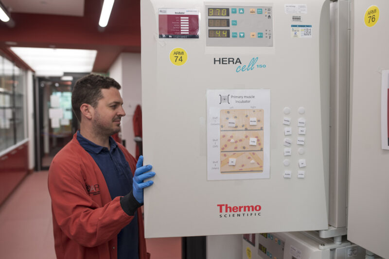
Now, Dr William Roman’s group at the Australian Regenerative Medicine Institute (ARMI) has launched their new imaging-based spatial transcriptomics platform, which dramatically reduces experimental costs from more than $35,000 down to approximately $30 per experiment, making spatial biology more accessible to researchers across Australia.
The new platform centres on a custom-built microscope system that combines sophisticated imaging capabilities with automated robotic technology. It employs a robotic arm to perform sequential imaging rounds, using precisely designed probes tagged with fluorophores to visualise RNA transcripts in their native spatial context.
Traditional sequencing-based spatial transcriptomics provides comprehensive genome-wide coverage but comes with prohibitive costs that limit experimental scope.
Dr Roman’s imaging-based approach offers a cost-effective solution for researchers who have identified specific gene panels of interest. Rather than paying tens of thousands for broad transcriptomic coverage, research groups can now focus on 50-100 key RNA signatures at a fraction of the cost, making longitudinal studies and large sample sizes financially viable.

“This is the perfect technique to use if you’ve already conducted bulk sequencing to identify your genes of interest, and now want to move on to characterising their role in the spatial context,” Dr Roman explained.
“You can explore how your genes are expressed in relation to each other, understand more about their relationship over time and within space, and discover unexpected spatial patterns.”
Understanding where genes are expressed in a tissue unlocks insights into everything, from embryo development to tumour architecture. Developmental biologists can track gene expression patterns during organ formation, whilst cancer researchers can examine tumour microenvironments and treatment responses. The platform particularly benefits researchers studying non-model organisms, such as various fish species for which commercial antibodies aren’t available.
With specialist postdoctoral researcher Dr Melinda Wang recently joining the team, the Roman Group is actively welcoming collaborative partnerships to explore new use-cases for the technology.
Explore a collaboration by contacting Dr William Roman here: william.roman@monash.edu
This article originally appeared on the ARMI website and was published here with permission.